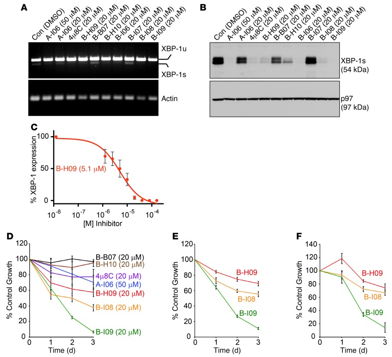
Figure 5. IRE-1 inhibitors with masked aldehyde moieties potently suppress XBP-1s expression and leukemic growth.
(A) WaC3 CLL cells were treated with indicated compounds for 24 hours and lysed for RNA extraction. Human unspliced XBP1 (XBP1u), spliced XBP1 (XBP1s), and actin were detected by RT-PCR using specific primers. Data are representative of 3 experiments. (B) Mouse B cells were stimulated with LPS for 48 hours to allow for XBP-1s expression, and then treated with indicated inhibitors for 24 hours. Lysates were immunoblotted for XBP-1s and p97. Data are representative of 3 experiments. (C) LPS-stimulated B cells were treated with 0, 1.25, 2.5, 5, 10, 20, 40, 80, or 160 mM B-H09 for 24 hours. Equal amounts of lysates were immunoblotted for XBP-1s. The XBP-1s protein band intensity was determined using ImageJ. The percentage of inhibition was determined by comparing with the untreated group. Data from 3 experiments were plotted as mean ± SEM. (D) Mouse CD3–IgM+CD5+ Eμ-TCL1 CLL cells were treated with DMSO or indicated inhibitors for 3 days and subjected to XTT assays. Percentages of growth were determined by comparing inhibitor-treated with DMSO-treated groups. Data from 4 identical experimental groups were plotted as mean ± SD. Results are representative of 3 experiments. (E and F) Primary CLL cells from 2 human patients were treated with DMSO or indicated inhibitors (20 μM), subjected to XTT assays, and similarly analyzed. Data from 4 identical experimental groups were plotted as mean ± SD. Results are representative of 3 experiments.

