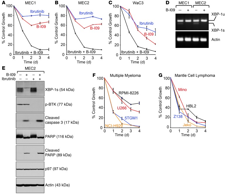
Figure 9. B-I09 synergizes with ibrutinib to induce apoptosis in human CLL, MM, and MCL cell lines.
(A–C) MEC1, MEC2 and WaC3 human CLL cells were treated with DMSO (control), B-I09 (20 μM), ibrutinib (10 μM), or a combination of both for 4 days, and subjected to XTT assays. Percentages of growth were determined by comparing inhibitor-treated groups with control groups. Data from 4 identical experimental groups were plotted as mean ± SD. Results are representative of 3 independent experiments. (D) MEC1 and MEC2 human CLL cells were treated with DMSO (control) or B-I09 (20 μM) for 48 hours and lysed for RNA extraction and RT-PCR. Human XBP1u, XBP1s, and actin were detected using specific primers. Results are representative of 3 independent experiments. (E) Human MEC2 CLL cells were treated for 72 hours with DMSO (control), B-I09 (20 μM), ibrutinib (10 μM), or the combination of B-I09 and ibrutinib. Cells were lysed for analysis of indicated proteins by immunoblots. Data are representative of 3 independent experiments. (F and G) MM cell lines (F) and MCL cell lines (G) were treated with DMSO or the combination of B-I09 (20 μM) and ibrutinib (10 μM) for a course of 4 days and subjected to XTT assays at the end of each day. Percentages of growth were determined by comparing treated groups with control groups. Each data point derived from 4 independent groups receiving exactly the same treatment was plotted as mean ± SD. Results are representative of 3 independent experiments.

