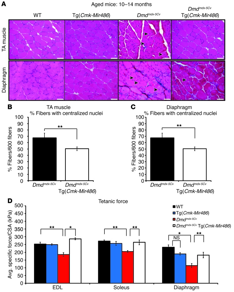
Figure 5. Aged dystrophic mice show similar improved pathology resulting from miR-486 transgenic overexpression.
(A) Representative H&E staining of TA and diaphragm muscles taken from aged (10 to 14 months old) WT, Tg(Cmk-Mir486), Dmdmdx-5Cv, and Dmdmdx-5Cv Tg(Cmk-Mir486) mice. Arrowheads represent centralized myonuclei. Scale bars: 50 μm. (B) Graphs of centralized myonuclei counts of the TA and (C) diaphragm muscles of aged Dmdmdx-5Cv and Dmdmdx-5Cv Tg(Cmk-Mir486) mice. Three mice (n = 6 TA and n = 3 diaphragm muscles) were used, with 600 TA and 200 diaphragm individual myofibers being counted. (D) Tetanic muscle-force measurements, normalized to CSA, of isolated EDL, soleus, and diaphragm muscles. Five (WT and Tg[Cmk-Mir486]) to 8 (Dmdmdx-5Cv and Dmdmdx-5Cv Tg[Cmk-Mir486]) aged (10 to 14 months old) male mice were used for each genotype cohort. *P < 0.005; **P < 0.05.

