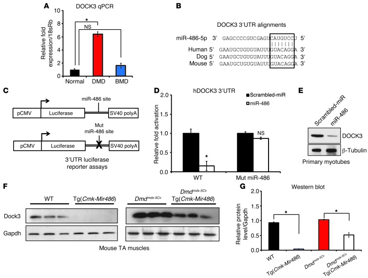
Figure 7. DOCK3 is a direct target of miR-486 in skeletal muscle.
(A) Real-time qPCR of human DOCK3 expression levels in normal, DMD, and BMD muscle biopsies. Expression levels are normalized to the 18sRb loading control. (B) Evolutionary conservation of the miR-486 seed site in mammalian DOCK3 3′ UTRs. Human, dog, and mouse are shown aligned with the seed region of miR-486-5p (boxed inset). (C) Schematic of 3′ UTR of the miR-486 target fused to a luciferase reporter construct. The miR-486 seed site is mutated in the mutant constructs. (D) Relative luciferase fold expression of the human DOCK3 3′ UTR fused to luciferase and transfected into HEK293T cells with either miR-486 or scrambled miR control plasmids. (E) Western blot analysis of human DOCK3 protein expression in primary human myotubes overexpressing either miR-486 or scrambled miR control lentivirus. β-Tubulin is shown as a loading control. (F) Western blot of DOCK3 protein in WT, Tg(Cmk-Mir486), Dmdmdx-5Cv, and Dmdmdx-5Cv Tg(Cmk-Mir486) TA muscle lysates. GAPDH is shown as a loading control. (G) Densitometry graph of DOCK3 protein expression normalized to either WT or Dmdmdx-5Cv levels,and the GAPDH loading control. *P < 0.005.

