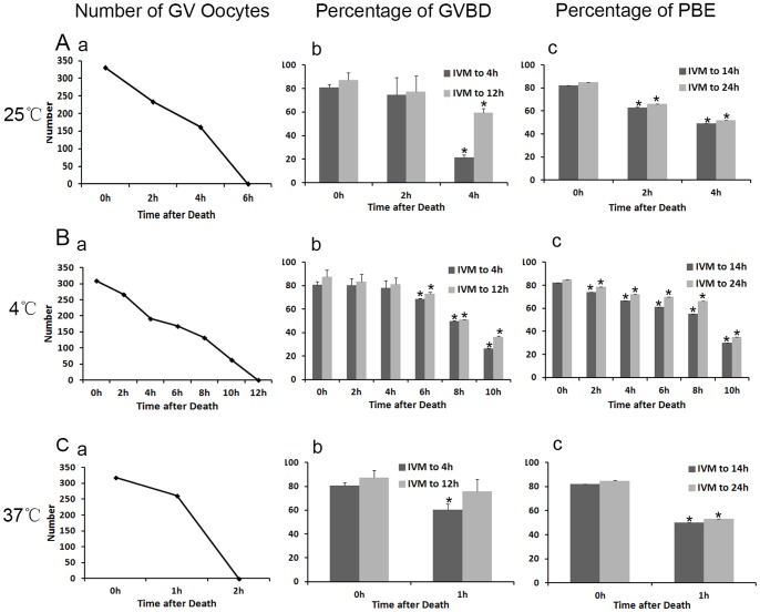
Figure 1. Effects of different carcass preservation periods on oocyte meiotic process.
(A) oocytes matured in vitro after carcass preserved for different times (0, 2 and 4 h) at room temperature (25°C). (a) The number of GV oocytes collected from the preserved carcass in the experimental group and control group. (b) The percentage of GVBD oocytes in the control group (n = 330), PMI 2 h group (n = 234) and PMI 4 h group (n = 162). Each bar represents mean ±SEM (n = 3). * indicate statistically significant differences (P<0.05). (c) The percentage of PBE in oocytes cultured in vitro in preservation group and control group. * P<0.05. (B a) The number of GV oocytes collected from the ovary after preservation at low temperature (4°C). (B b) The percentage of GVBD oocytes in the control group (n = 309) and the preservation group (n = 267, 192, 168,132 and 63). * P<0.05. (B c) The percentage of PBE in oocytes cultured in vitro in different groups. * P<0.05. (C) The number of GV oocytes, percentage of GVBD oocytes and PBE same as A at high temperature (37°C). * P<0.05.

