
Figure 3. Morphological analysis of reproductive organs from worms treated with dsSmvkr1 or dsSmvkr2 RNA.
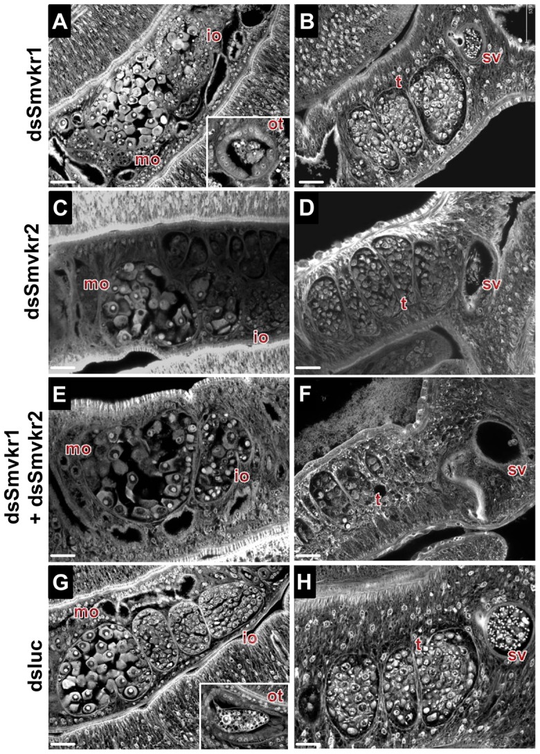
CLSM images of whole-mount preparations of S. mansoni worm couples stained with carmine red. Worms were treated exactly as described in Fig. 2 with dsSmvkr1 (A, B), dsSmvkr2 (C, D), dsSmvkr1 and dsSmvkr2 (E, F) or control dsLuc (G, H) RNA. The morphology of female (left) and male (right) reproductive organs was analyzed. io: immature oocytes, mo: mature oocytes, ot: ootype, sv: sperm vesicle, t: testes. Scale bar: 20 µm.