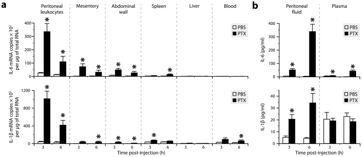
Figure 1. Both IL-6 and IL-1β are produced in tissues exposed to PTX, but only IL-6 reaches increased levels in the circulation.
a, Quantification of the mRNAs encoding IL-6 and IL-1β by qRT-PCR in different tissues from mice killed 3 or 6 h after intraperitoneal injection of PTX (20 µg/kg) or PBS. *Significantly different from the corresponding PBS group according to the Wilcoxon test (P<0.05). Sample size: 4–10 (PBS groups) or 7–10 (PTX groups). b, Quantification of IL-6 and IL-1β by ELISA in peritoneal fluid and plasma samples. *Significantly different from the corresponding PBS group according to the Wilcoxon test (P≤0.0003). Sample size: 3–9 (PBS groups) or 4–10 (PTX groups).

