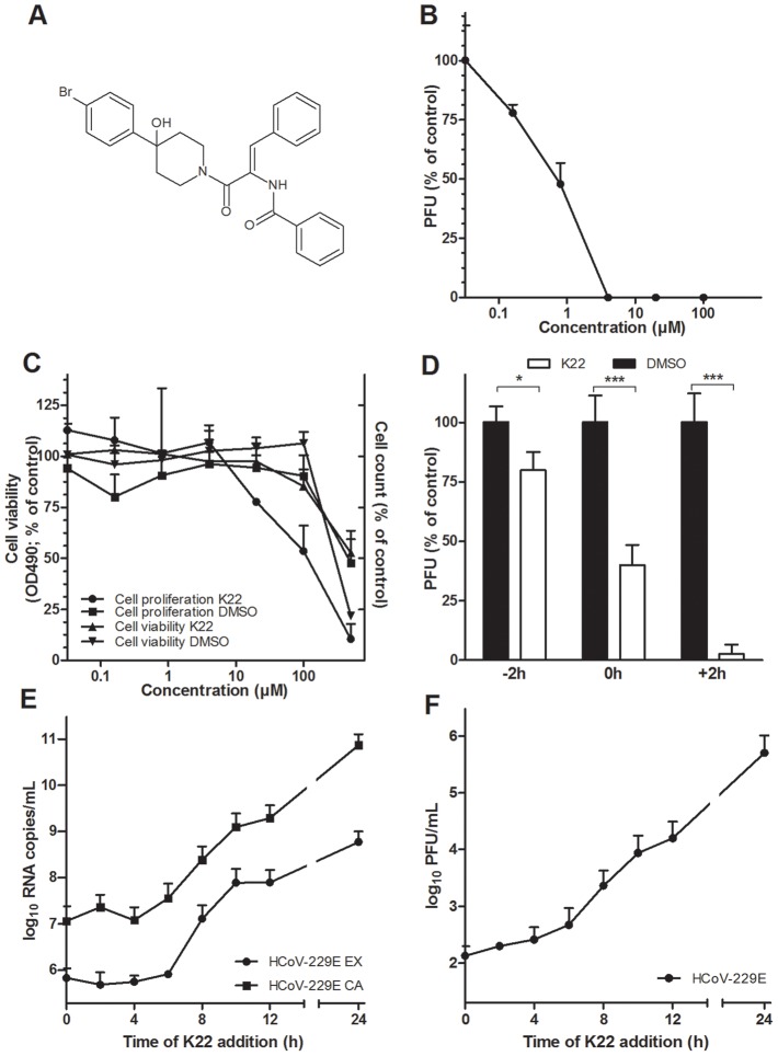
Figure 1. K22 structure, antiviral activity, and cytotoxicity.
(A) K22 structure. (B) Anti-HCoV-229E activity of K22 in MRC-5 cells. K22 and HCoV-229E were added to MRC-5 cells, and the number of viral plaques developed after 48 h were assessed. Data shown are means (±SD) of duplicate determinations from three independent experiments. PFU, plaque forming unit. (C) Viability and proliferation of MRC-5 cells in the presence of K22. MRC-5 cells were incubated with K22 or DMSO solvent for 48 h at 37°C and the cell viability determined using tetrazolium-based reagent while cell proliferation was assayed by counting of cells. Data shown are means (±SD) of duplicate determinations from two independent experiments. (D) K22 affects the post-entry phase of viral life cycle. K22 (4 µM) or DMSO solvent were incubated with cells for a period of 2 h either before (−2 h), during (0 h) or after a 2 h period of cell inoculation with HCoV-229E, and the number of viral plaques developed after 48 h were assessed. Data shown are means of duplicate determinations from three independent experiments.*P<0.05; n = 3. ***P<0.005; n = 3. (E-F) K22 exhibits potent antiviral activity when added up to 6 h after infection of cells. MRC-5 cells were inoculated with HCoV-229E at a moi of 0.05 for 45 min at 4°C, and K22 (10 µM) added at specific time points relative to the end of inoculation period. The culture medium and cells were harvested after 24 h of incubation at 37°C, and the viral RNA (E) and infectivity (F) determined. Data shown are means (±SD) of duplicate determinations from two independent experiments. EX, extracellular medium; CA, cell-associated sample.

