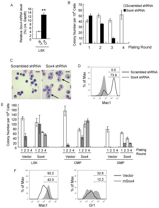
Figure 1. Sox4 is required for abnormal serial-replating ability and myeloid differentiation block of Cebpa KO stem/progenitor cells.
(A) Sox4 expression was analyzed by qPCR in LSK cells (Lin−c-kit+Sca1+) sorted from Mx1-Cre−CebpaloxP/loxP (WT) and Mx1-Cre+ CebpaloxP/loxP (KO) mice 14 days after polyI:C injection. Relative gene expression levels were determined as % Gapdh.
(B) Cebpa KO LSK cells were transduced with lentiviruses harboring either scrambled shRNAs or Sox4 shRNAs and replated in methylcellulose plus puromycin. The bar chart shows the colony number for four rounds.
(C) Wright-Giemsa staining of cells from the first plating round in Figure 1B. Data are representative of 3 independent experiments. Macrophages are indicated with arrows. Scale bar, 20 μm.
(D) Flow cytometry analysis of cells from the first plating round in Fig 1B. Shadow histogram indicates scrambled shRNA-infected cells, and black line indicates Sox4 shRNA-infected cells. Percent cells are shown for the indicated gates. Data is representative of 3 independent experiments.
(E) Wild-type LSK, CMP (Lin−c-kit+Sca1−CD34+FcRgII/IIIlo) and GMP (Lin−c-kit+Sca1−CD34+FcRgII/IIIhigh) were transduced with an empty retrovirus (Vector) or a retrovirus expressing Sox4 (Sox4) and replated as Figure 1B.
(F) Wild-type LSK cells were transduced as in Figure 1E and grown in liquid culture plus puromycin and cytokines. 7 days later, Mac1 (left) and Gr1 (right) expression was analyzed by flow cytometry. Shadow histograms indicate cells infected with empty virus, and black lines indicate cells overexpressing Sox4. Percent cells are shown for the indicated gates. Data are representative of 3 independent experiments. Error bars indicate the mean ± SEM of 3 independent experiments. **: p <0.001. See also Figure S1 and Table S1.

