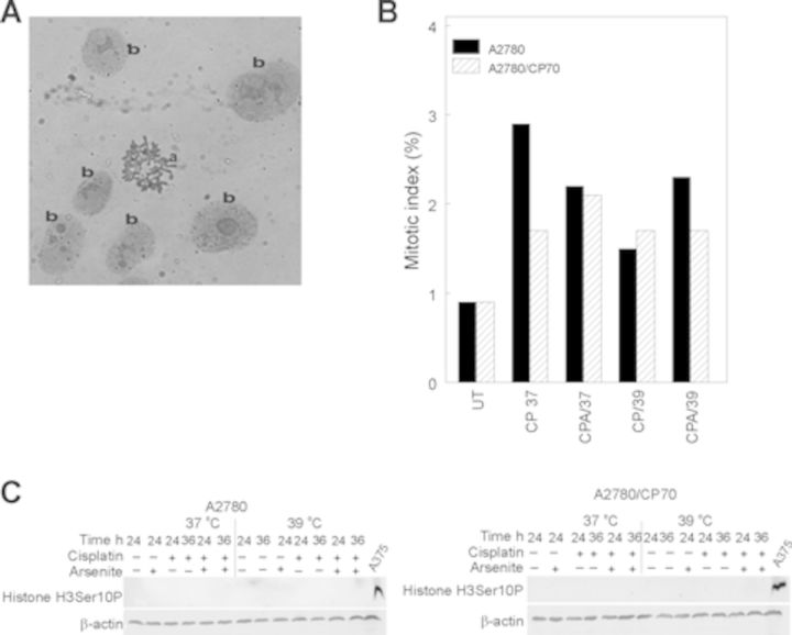
FIG. 3.
Mitotic index determination and Western blot analysis of protein marker of mitosis. (A) Representative picture of (a) mitotic spread and (b) interphase nuclei. (B) Plot of means of percent mitotic cells for duplicate slides. Cells were treated with their respective IC50 cisplatin (CP) (A2780, 4 μM; CP70, 40 μM) or CP plus 20 μM sodium arsenite (CPA) at 37 or 39°C (hyperthermia) for 1 h. Treated cells were washed with PBS and refed fresh media and incubated at 37°C for 36 h. Mitotic index was determined at 36 h after treatment. Data are single biological experiments performed in duplicate dishes. C. Western blot analysis of histone H3Ser10P. Protein lysates were prepared 36 h after treatment for Western blot analysis of histone H3Ser10P. Data are representative from duplicate biological experiments. A375 human melanoma cells were treated with 5μM sodium arsenite for 24 h served as positive control for mitotic cells (McNeely et al.,2008b). ß-actin served as loading control.

