Figure 1.
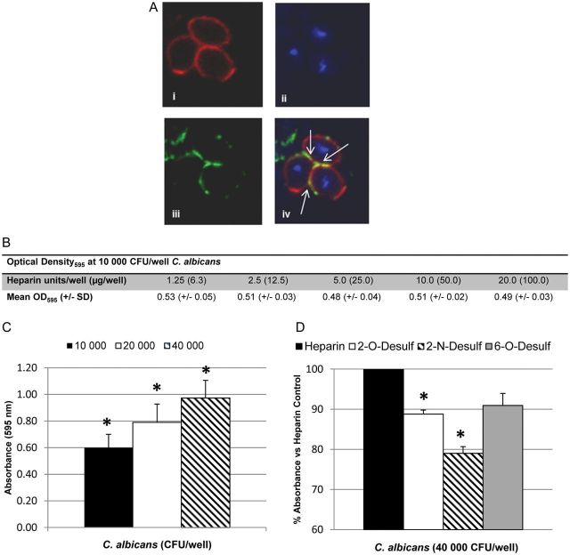
Candida albicans binds heparin in vitro. A, Confocal microscopy of C. albicans after staining with PKH26 (top left), 4′,6-diamidino-2-phenylindole dihydrochloride (top right), or heparin–Alexa Fluor 488 (bottom left). Arrows (bottom right) show colocalization of heparin with C. albicans cell surface. B, Binding of 10 000 colony-forming units (CFU) of C. albicans (optical density at 595 nm [OD595]) to increasing concentrations of heparin immobilized on an allylamine-coated 96-well microtiter plate. Values represent means±standard deviations (SDs) of duplicate wells. C, Heparin-binding enzyme-linked immunosorbent assay with 2.5 U of heparin per well and increasing C. albicans input. Graph represents means ± SDs of 4 experiments; *P < .007 for all inputs. D, Binding of C. albicans (40 000 CFU per well) to equimolar amounts of heparin analogs desulfated at the 2-O, 2-N, or 6-O positions versus heparin control (normalized to 100%). Graph represents means ± SDs of 3 experiments, performed in triplicate *P ≤ .003 vs heparin control.

