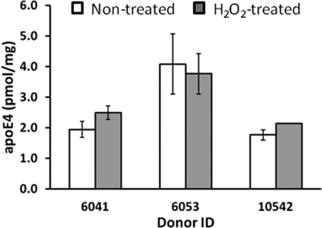
Figure 3.
Oxidation of Met in the LGADMEDVR peptide with H2O2. Measurements were performed on the temporal cortex from severe AD patients (donor IDs are 6041, 6053, and 10542, Supporting Information, Table S1). For quantification of apoE4 in nontreated samples, the transitions for LGADMEDVR were used. For quantification of apoE4 in H2O2-treated samples, the transitions for LGADM(O2)EDVR were used (Table S2, Supporting Information). The concentration was calculated for three experimental replicates by monitoring three transitions per individual peptide and is presented as the mean ± SD.

