Fig. 2.

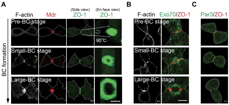
Assembly and opening of a disc-shaped tight junction at the division site is associated with de novo bile canaliculus formation. (A) Localization of F-actin (gray), Mdr (red) and ZO-1 (green) during different stages of bile canaliculus (BC) formation in post-cytokinesis cells. All images are snapshots of 3D reconstructions (0.5-µm×8–10 optical slices) at the indicated angles (side or en-face). Pre-bile-canaliculus, small bile canaliculus, and large bile canaliculus stages were defined as those displaying a single line, two lines separated by a short space, and two puncta of ZO-1 signal (side view), respectively. (B) Exo70 (green) localization with respect to ZO-1 (red) and F-actin (gray) during different stages of bile canaliculus formation. (C) Colocalization of Par3 (green) and ZO-1 (red) during different stages of bile canaliculus formation. Dotted lines denote the cell outline. Scale bars: 3 µm.
