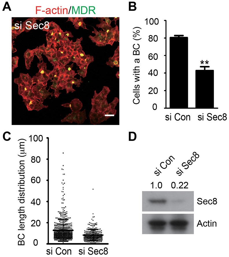
Fig. 5.

The exocyst is required for bile canaliculus formation. (A) Representative image of bile canaliculus formation in the Sec8-knockdown (si Sec8) cells. F-actin, red; Mdr, green. (B,C) The percentage of cells that were engaged in bile canaliculus formation (B) and bile canaliculus length distribution (C) in the control (the same cells as those shown in Fig. 4A) and si Sec8 cells were quantified. (D) Western blotting indicated that the level of Sec8 (relative molecular mass: 110 kD) in the si Sec8 cells was reduced to 22% of that in the control cells (the figures are shown across the top of the blot). Actin was used as a loading control. Scale bar: 10 µm.
