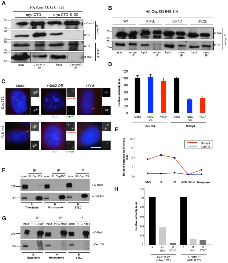
Fig. 7.
C-Nap1 phosphorylation disturbs its interaction with Cep135 in mitosis. (A) HA–Cep135-CTD (648–1141) was co-transfected with Myc–C-Nap1-CTD or CTD-S10D into U2OS cells. Cell lysates were immunoprecipitated (IP) using antibodies against Myc or HA, and western blotted with antibodies against C-Nap1, Myc or HA, as indicated. Note that in the anti-Myc blot, shorter exposures are shown for the inputs, but exposures for the two IPs are the same. (B) As for A, except using the Myc–C-Nap1-CTD constructs indicated and immunoprecipitations were only performed with anti-Myc antibodies. WT, wild type. (C) U2OS cells were mock treated, transfected for 24 hours to overexpress Nek2 (Nek2 OE) or stimulated with EGF for 4 hours before being fixed and stained with antibodies against γ-tubulin (red) and Cep135 or C-Nap1 (green), along with the DNA stain Hoechst 33342 (blue). Scale bar: 5 µm. (D) Centrosome intensities of Cep135 and C-Nap1 in the cells overexpressing Nek2 or treated with EGF were determined relative to the mock-treated cells. a.u., arbitrary units. Data show the mean±s.d. (three independent experiments). (E) HeLa cells were synchronized with a double-thymidine block and released for 0 hours (G1/S), 4 hours (S) or 9 hours (G2). Metaphase and telophase cells were identified in an unsynchronized population by chromosome status. In each case, cells were fixed and stained with Cep135 and C-Nap1 antibodies, and cell cycle status was confirmed by staining for cyclin B1. Centrosome intensities were measured and are presented as means relative to that of metaphase cells (n = 50). (F,G) HEK 293T cells that were arrested in the cell cycle as indicated were lysed and immunoprecipitated using pre-immune serum (PI) and anti-Cep135 (F) or anti-C-Nap1 (G) antibodies. Lysates (input) and immunoprecipitates were western blotted with antibodies against Cep135 and C-Nap1, as indicated. Molecular mass (kDa) is shown on the left of panels A, B, F and G. (H) Quantification of data from F and G showing the extent of co-precipitation in mitotically-arrested cells relative to that seen in S-phase-arrested cells. Noc, nocodazole; WB, western blotted.

