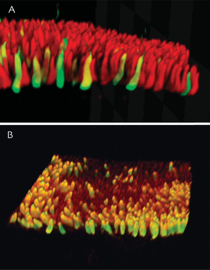
Fig. 3.
Examples of hair cells in vestibular end organs of the toadfish. All hair cells in anamniotes such as the toadfish have Type II hair cell-like structure and connectivity. A: Volume rendering of an image stack through a portion of the toadfish horizontal canal crista ampullaris immunostained for glutamate (red) and GABA (green). B: Phalloidin (red) and GABA (green) immunostaining of the utricular macula. Both images were acquired using multiphoton microscopy, and the image stacks were rendered and rotated.

