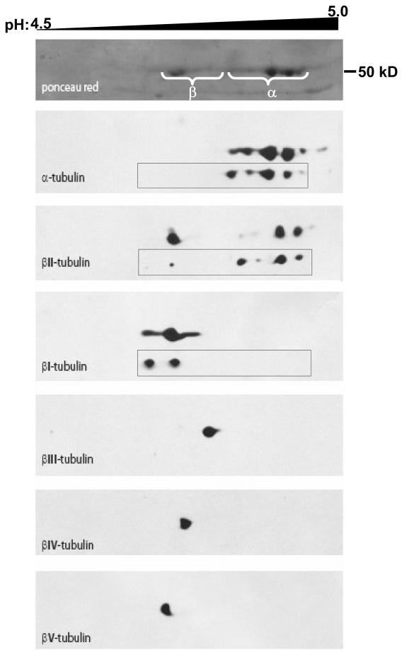
Figure 3. High resolution 2D-electrophoresis tubulin isotypes in Taxol-stabilized microtubules from HMEC-1, endothelial cells.
Tubulin was isolated from HMEC-1 cells as described in the text. Tubulin isotypes were separated in the first dimension on a 24 cm IPG-strip pH 4.5–5.5 (pH 4.5–5.0 portion shown). The second dimension was run on a 10% acrylamide gel and proteins were blotted on a nitrocellulose membrane. The membrane was probed with antibodies against the indicated isotypes and stripped between each antibody. Alignment of blots was performed using the actin spots (not displayed). An anti-human βV-tubulin rabbit polyclonal antibody was produced and specifically labeled a spot on the acidic side of βI-tubulin as predicted. Insets show equivalent blots for α-, βI- and βII-tubulins in A549 cells; the most acidic spot for βI-tubulin corresponds to its monoglutamylated form.

