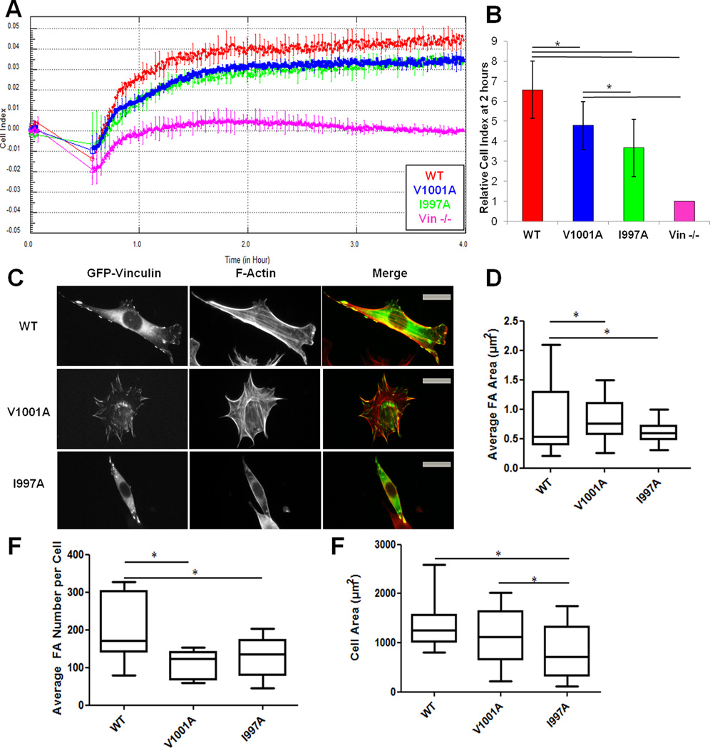
Figure 2. Vcn variants deficient in actin binding affect spreading and cell adhesion in MEFs.
(A) RTCA using the xCELLigence system shows that Vin−/− cells expressing VcnWT have higher cell impedance, hence more spread, than cells expressing VcnV1001A, VcnI997A or Vin−/−.MEFs. A representative trace of cell impedance (graphed as cell index (CI)) taken every 15 seconds for 13 hours; lower impedance indicates less contact with the sensor. Each data point represents an average CI of at least triplicate wells for each condition. (B) A graph showing the relative CI of cells spread on FN two hours following plating, which corresponds to the same time as the pictures shown in (C). Data is the average ± SEM combined from four independent experiments. *p≤ 0.05, in comparison to VcnWT. (C) Vin−/− MEFs transfected with GFP-tagged VcnWT, VcnI997A, or VcnV1001A and plated on FN for two hours. VcnI997A and VcnV1001A exhibit the same localization as VcnWT. (D,E,F) Box and whisker plots of FA area (D), FA number (E) and cell area (F). Areas were calculated using Matlab (Methods) (n=25). Cells expressing VcnI997A and VcnV1001A had fewer and larger FAs, *p-value≤0.05. Cells expressing VcnI997A were significantly smaller, but those expressing VcnV1001A were not. Scale bar is 25 µm.

