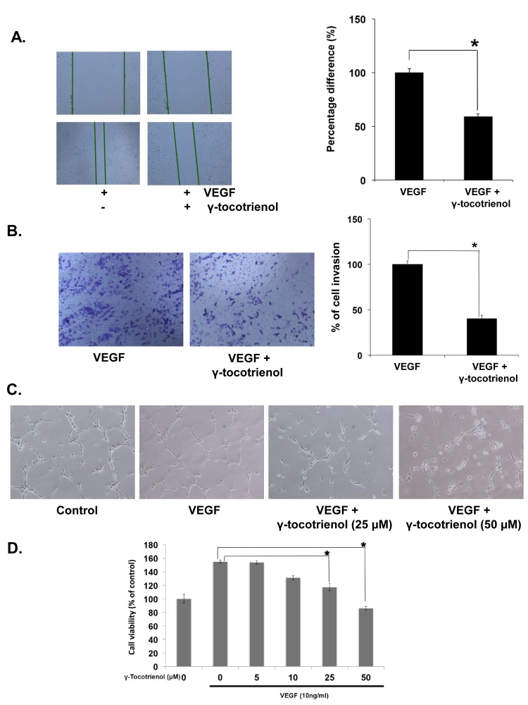
Figure 1. γ-tocotrienol inhibits VEGF-induced endothelial cell migration, invasion, capillary structure formation in vitro and cell viability.
A, γ-tocotrienol inhibited HUVEC migration. An IBIDI culture insert (IBIDI GmbH) consists of two reservoirs separated by a 500 μm thick wall created by a culture insert in a 35mm petri dish. An equal number of HUVECs (70 μl; 5×10⁵ cells/ml) were added into the two reservoirs of the same insert and incubated at 37°C/5% CO2. After 12 hours, the insert was gently removed creating a gap of ~500 μm. The cells were treated with 50 μM γ-tocotrienol for 12 h before being exposed to 10ng/mL VEGF for 24 h. Width of wound was measured at time zero and 24h of incubation with and without γ-tocotrienol. The representative photographs showed the same area at time zero and after 24 h of incubation. B, γ-tocotrienol inhibited HUVEC invasion through matrigel coated polycarbonate membrane. After pre-incubation with or without 50 μM γ-tocotrienol for 12 h, transwell chambers were then placed into the wells of a 24-well plate, in which we had added Medium 200 containing 10 ng/mL VEGF. After incubation for 24h cell invasion was analyzed and columns represent mean number of invaded cells. C, γ-Tocotrienol inhibited the VEGF-induced tube formation of endothelial cells in matrigel. After incubation, endothelial cells were fixed, and tubular structures were photographed (magnification, ×100). D, γ-Tocotrienol significantly inhibited the VEGF-induced cell survival of HUVECs. Cell viability was determined by MTT assay. *, p < 0.01 versus VEGF alone.

