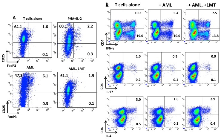
Figure 4. Effects of IDO-Expressing AML Blasts on T-Cell Expression of FoxP3 and on Cytokine Production Profile.
AML blasts were pre-treated for 2 hours with 200 μM 1MT or were left untouched, followed by stimulation with 100 ng/ml IFN-γ. IDO-expressing AML blasts were then co-cultured with allogeneic naïve CD4+ T cells in a standard mixed tumor lymphocyte culture (MTLC). (A) After 5 days in MTLC, cells were harvested, fixed, permeabilized and labeled with anti-CD25 and anti-FoxP3 mAbs, as detailed in Materials and Methods. Control cultures were established with CD4+ T cells and IL-2, either alone or combined with PHA as a polyclonal stimulus. Similar results were obtained in 3 independent experiments. The percentage of cells staining positively for a given antigen is indicated; (B) After 5 days in MTLC, T cells were activated for 6 hours with a polyclonal stimulus, consisting of PMA and ionomycin, in the presence of brefeldin-A to inhibit intracellular protein transport. Cells were then fixed, permeabilized and labeled with cytokine-specific mAbs, as detailed in Materials and Methods. The percentage of cells staining positively for a given antigen is indicated. Similar results were obtained in 3 independent experiments.

