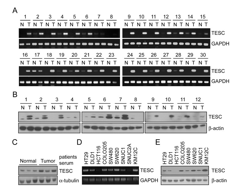
Figure 1. Increased mRNA and protein expression of TESC in human colorectal tissues, serum, and various CRC cell lines.
(A) RT-PCR analysis of TESC expression in 30 paired samples of non-tumor colon (N) and colorectal cancer (T) tissues. GAPDH was used as the loading control. (B, C) Western blot analysis of TESC expression in 12 pairs of non-tumor (N) and tumor (T) tissues (B), and in sera from patients with CRC (C). β-actin or α-tubulin was used as the loading control. (D, E) TESC expression in 10 CRC cell lines by RT-PCR (D) and Western blot (E) analysis.

