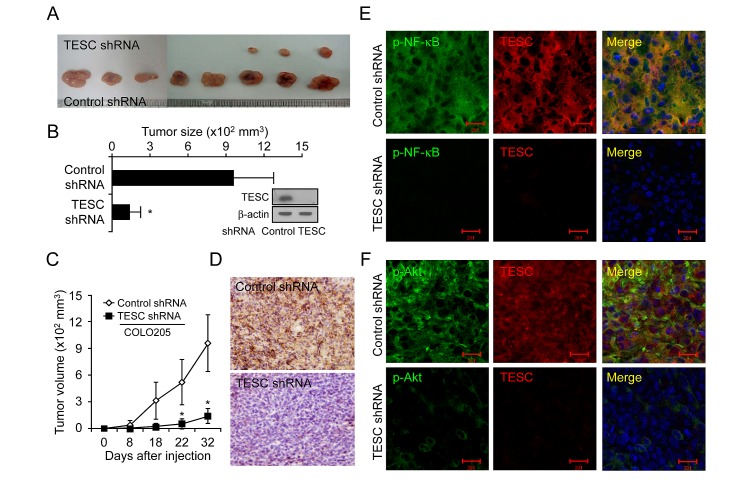
Figure 6. Suppression of TESC markedly reduces tumor growth in a CRC xenograft model.
(A-C) COLO205 cells transfected with control shRNA or TESC shRNA were inoculated into the right flank of 6-week-old nude mice. Tumor growth was monitored on the indicated days. Results represent mean tumor volume ± SD for eight animals; *P < 0.01 vs. control shRNA group. (D) Xenograft tumors formed by COLO205 cells expressing control shRNA or TESC shRNA were sectioned and stained for TESC expression. Bar = 50 μm. (E, F) Subcellular localization of TESC and phospho-NF-κB or phospho-Akt in xenograft tumors formed by COLO205 cells expressing control shRNA or TESC shRNA was detected with Alexa 555-conjugated secondary antibodies for TESC and Alexa 488-conjugated phospho-NF-κB or phospho-Akt antibodies. Bar = 20 μm.

