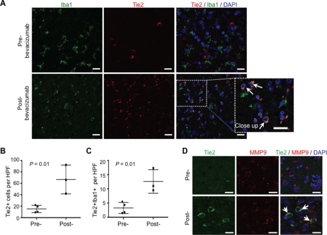
Figure 5. TEMs can be detected in human surgical glioblastoma specimens after bevacizumab therapy.
(A) Glioblastoma specimens after treatment with standard chemotherapy (pre-) or with bevacizumab (post-) were analyzed for Iba1 (green) and Tie2 (red) expression. DAPI was used for nuclear staining (blue). Scale bars = 20 μm. Note the over-representation of TEMs (Tie2+Iba1+ cells) in the post-bevacizumab tumor. White arrows indicate the presence of Tie2+Iba1+ cells. Scale bars = 20 μm. (B) Quantification of Tie2+ cells in surgical human glioblastoma recurring after standard therapy (Pre-; n = 4) or after bevacizumab (Post-; n = 3). Data are represented as mean ± SD of Tie2+ cells present in a HPF. (C) Quantification of Tie2+Iba1+ double positive cells in surgical human glioblastoma recurring after standard therapy (Pre-; n = 4) or after bevacizumab (Post-; n = 3). Data are represented as mean ± SD of Tie2+ cells present in a HPF. (D) Tie2+ cells overexpressed MMP9, as indicated with the double immunofluorescence for Tie2 (green) and MMP9 (red) expression. DAPI was used for nuclear staining (blue). White arrows indicate the presence of Tie2+MMP9+ cells. Scale bar = 20 μm.

