
Figure 1. Ectopic expression of c-kit in ACC-M cells induced an EMT program.
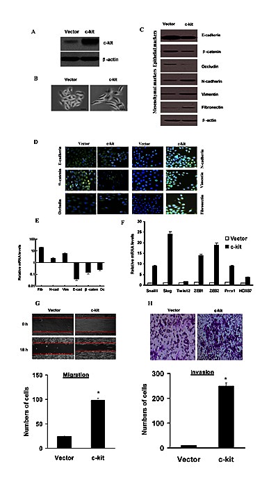
A), Immunoblotting assessment of the ectopic c-kit protein expression after plasmid transfection in ACC-M cells. B), Morphologic change of ACC-M cells expressing c-kit or empty vector. Scale bar, 100 mm. C), Immunoblotting analysis of expression of the epithelial markers E-cadherin, β-catenin, and Occludin, and the mesenchymal markers Fibronectin, Vimentin, and N-cadherin. D), Immunofluorescence staining for the epithelial and mesenchymal markers. Scale bar, 100 mm. E), The expression of E-cadherin, β-catenin, Occludin, fibronectin, Vimentin, and N-cadherin mRNAs were assessed by real-time PCR. F), mRNA expression levels of known EMT inducers were assessed by real-time PCR. Error bars represent the mean±SD of triplicate experiments. G and H), Migration (G) and invasion (H) assays in stable ACC-M cells. The mean was derived from cell counts of 5 fields, and each experiment was repeated 3 times (* P < 0.001, compared with the control). Representative images of migrated and invaded cells are shown.