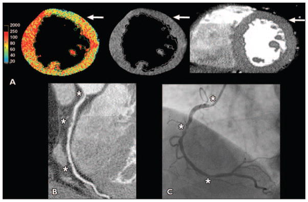
Fig. 4. 63-year-old obese man with sedentary lifestyle who presented with atypical chest pain (patient 2 in Table 3).

A, Color and gray-scale CT–myocardial perfusion imaging showed anterior artifacts where hypoperfusion areas did not cross phase boundaries (arrows), but no myocardial regions had defined hypoperfusion.
B, Cardiac CT showed right coronary artery ostial 70% stenosis, mid maximal 90% stenosis, and distal 70% stenosis (asterisks) as well as left anterior descending artery and ramus stenoses of ≤ 50%.
C, Patient was ruled in for myocardial infarction, and invasive angiography also showed stenoses (asterisks); he received two right coronary stents.
