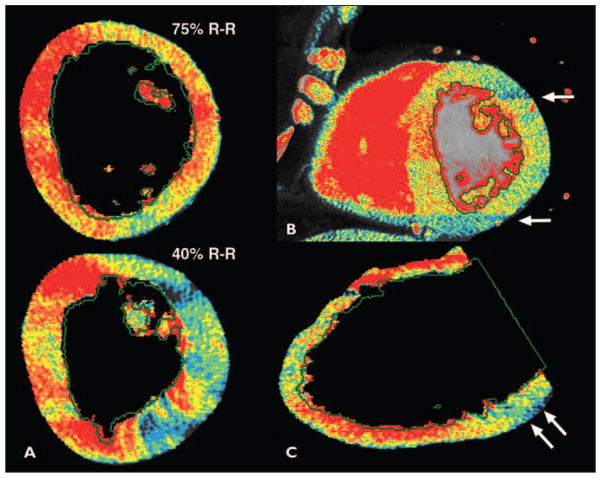
Fig. 5. Common artifacts with CT–myocardial perfusion imaging (MPI).
A, Variations in CT–myocardial perfusion imaging (MPI) over cardiac R-R cycle, or beat-to-beat variability, may result in either false-positive or false-negative findings; 40% R-R interval image was false-positive.
B, CT-MPI defects that are adjacent to phase-determined boundary, but that fail to cross phase-determined boundary (arrows), are deemed artifact.
C, Inferolateral defect due to beam-hardening from spine or descending aorta.

