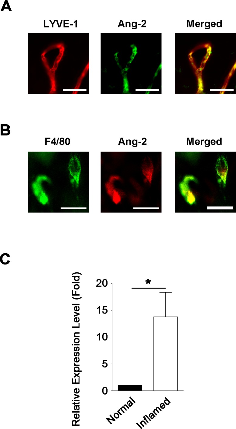
Figure 1.

Angiopoietin-2 is expressed on lymphatic vessels and macrophages in inflamed cornea. (A) Representative images of immunofluorescent microscopic assays showing Ang-2 (green) coexpressed with LYVE-1+ (red) lymphatic vessels in the inflamed cornea. Scale bar: 100 μm. (B) Representative images showing Ang-2 (red) expression on F4/80+ (green) macrophages in the inflamed cornea. Scale bar: 10 μm. (C) Quantitative PCR analysis showing significant increase of Ang-2 expression in corneal macrophages in the inflamed condition. *P < 0.05.
