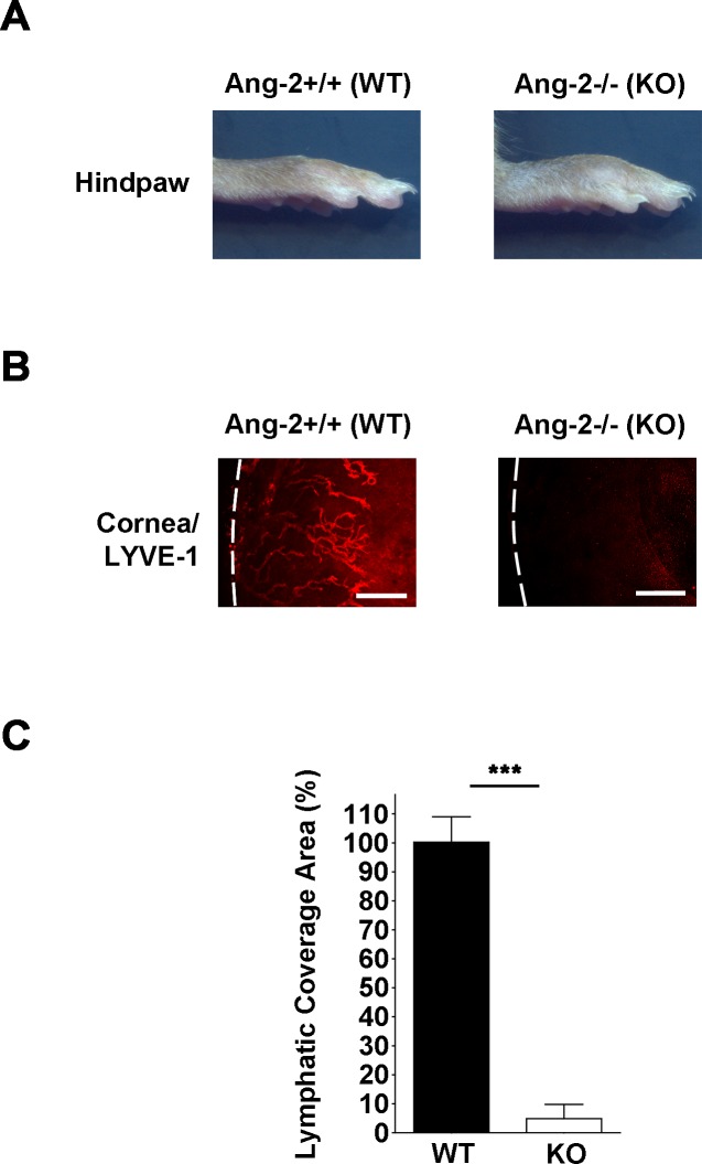
Figure 2.
Corneal LG response is inhibited in Ang-2 knockout mice. (A) Angiopoietin-2 knockout mice showing swollen hind paws reflecting lymphedema. (B) Representative images of immunofluorescent microscopic assays showing significantly reduced lymphatic vessels (LYVE-1+, red) in the inflamed cornea of Ang-2 knockout mice compared with wild-type controls. Corneas were harvested 14 days after suture placement. White dotted line: demarcation between the cornea and conjunctiva. Scale bars: 200 μm. (C) Summarized data from repetitive experiments showing significant difference in lymphatic invasion area between the two groups. ***P < 0.001.

