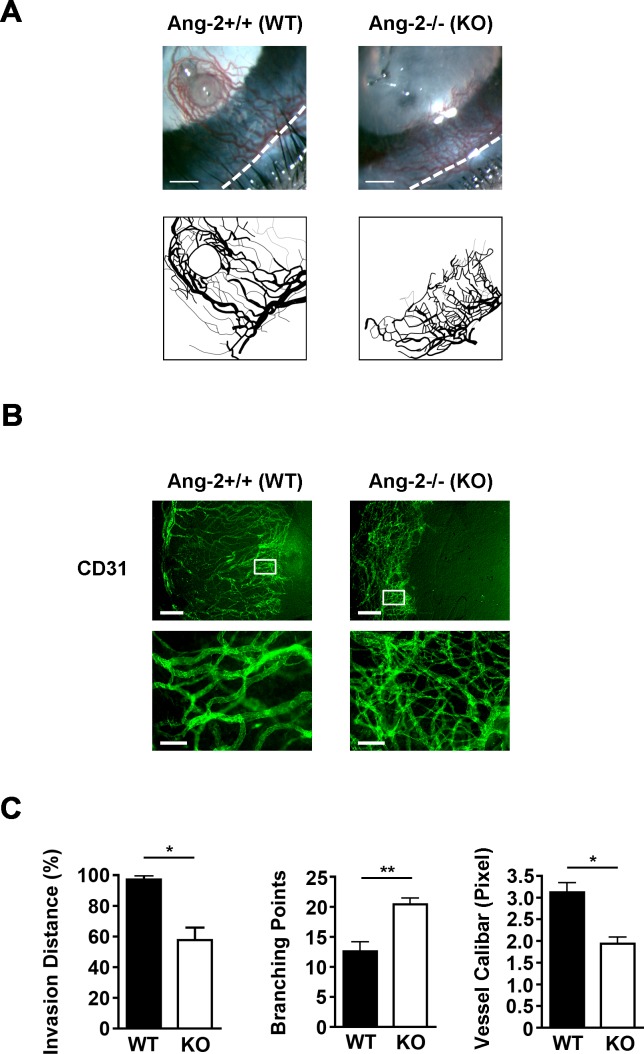
Figure 3.
Abnormal patterning of blood vessels in Ang-2 knockout mice. (A) Representative images from ophthalmic slit-lamp bioscopy showing disorganized and shortened blood vessels in Ang-2 knockout mice. White dotted line: demarcation between the cornea and conjunctiva. Scale bar: 350 μm. (B) Representative images of immunofluorescent microscopic assays showing difference of blood vessels in inflamed corneas of Ang-2 and wild-type control mice. Scale bar: 250 μm. Bottom panels: higher magnification view of boxed areas in the upper panels showing an increase in branching points and a decrease in diameters of blood vessels in Ang-2 knockout mice. Scale bar: 100 μm. (C) Summarized data from repetitive experiments showing the differences of blood vessels in Ang-2 knockout and wild-type mice in terms of invasion area, branching point, and diameter. *P < 0.05. **P < 0.01.

