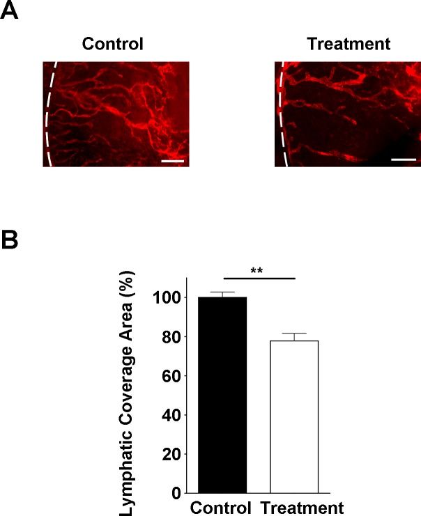
Figure 5.
Corneal LG response is inhibited by anti-Ang-2 treatment. (A) Representative images of immunofluorescent microscopic analysis showing significantly reduced lymphatic vessels (LYVE-1+, red) in the inflamed cornea of Ang-2 siRNA treatment group. White dotted line: demarcation between the cornea and conjunctiva. Scale bar: 200 μm. (B) Summarized data from repetitive experiments showing significant difference in lymphatic invasion area. **P < 0.01.

