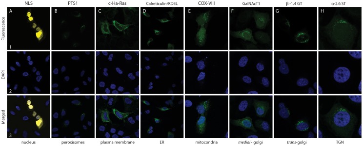
Figure 5. Confocal laser microscopy of U-2-OS cells expressing localized fluorescent proteins.
Confocal laser microscopy of fixed U-2-OS cells transiently expressing fluorescent proteins localized to major cellular compartments. Shown are representative images of eYFP or eGFP detected 48 hours after transfection: (A1-3) pFAST6-eYFP::NLS, (B1-3) pFAST5-eGFP::PTS1, (C1-3) pFAST37-eGFP::c-Ha-Ras, (D) pFAST57-CRT::eGFP::KDEL, (E) pFAST58-COXVIII::eGFP, (F) pFAST59-GalNAcT1::eGFP, (G) pFAST61-b1,4GT::eGFP, (H) pFAST56-a-2,6ST-eGFP. (A1–H1) microscopy with fluorescence filters. (A2–H2) nuclei stained with DAPI (dark blue). (A3–H3) merged pictures.

