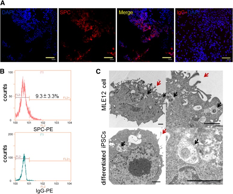
Figure 2.
Evaluation of differentiated iPSC phenotypes on day 12. (A): iPSCs differentiated for 12 days were immunostained with goat anti-mouse pro-SPC antibody (red) or goat IgG and nuclear counterstained with DAPI (blue). Scale bars = 50 μm. (B): Flow cytometry analysis of SPC expression. (C): Transmission electron micrographs of MLE12 cells (a murine type 2 pneumocyte cell line) and differentiated iPSCs. Upper left: MLE12 cells exhibit characteristic lamellar bodies (black arrows) and apical microvilli (red arrows). Upper right: Magnified view of the upper-left image. Lower left: Mouse differentiated iPSCs showing similar lamellar bodies and microvilli to those of MLE12 cells. Lower right: Magnified view of the lower-left image. Scale bars = 1 μm. Abbreviations: DAPI, 4′,6-diamidino-2-phenylindole; iPSC, induced pluripotent stem cell; PE, phycoerythrin; SPC, surfactant protein C.

