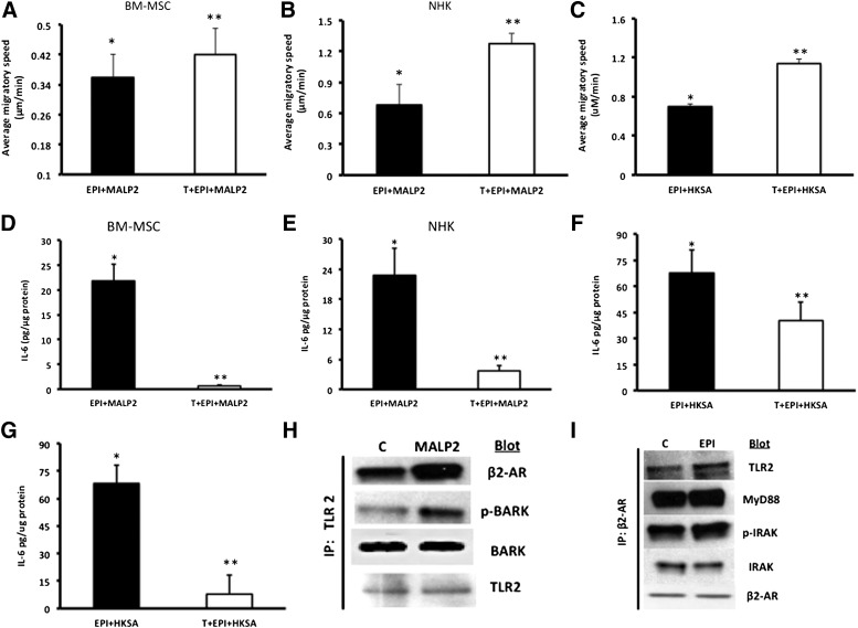
Figure 4.
Timolol (a nonselective β2-AR antagonist) reverses the combined effects of EPI + MALP2 or EPI + HKSA on single-cell migration (SCM) and IL-6 secretion in BM-MSCs and NHKs. (A): BM-MSCs were exposed to EPI (50 nM) + MALP2 (100 ng/ml) or pretreated with Timolol (10 μM, 30 minutes) and were further treated for 4 hours with EPI + MALP2. SCM rates of at least 50 cells per treatment were determined, as described in Materials and Methods. Values are expressed as average migratory speed (μm/min; mean ± SD). ∗, p < .05 versus EPI or MALP2; ∗∗, p < .005 versus EPI + MALP2 (n = 3 experiments; total of 150 cells). (B, C): NHKs were exposed to EPI (50 nM) + MALP2 (100 ng/ml), EPI + HKSA (104 cells per milliliter), or pretreated with Timolol (10 μM, 30 minutes) and were further treated for 4 hours with EPI + MALP2 or EPI + HKSA. SCM rates of at least 60 cells per treatment were determined, as described in Materials and Methods. Values are expressed as average migratory speed (μm/min; mean ± SD). ∗, p < .05 versus EPI or MALP2; ∗∗, p < .05 versus EPI + MALP2; ∗∗, p < .01 versus EPI + HKSA (n = 2 experiments; 120 cells). (D, F): BM-MSCs were exposed to EPI (50 nM) + MALP2 (100 ng/ml), EPI (50 nM) + HKSA (104 cells per milliliter), or pretreated with Timolol (10 μM, 30 minutes) and were further treated for 4 hours with EPI + MALP2 or EPI + HKSA. Secreted IL-6 in the cell culture supernatant was determined using enzyme-linked immunosorbent assay, as described in Materials and Methods. Values are expressed as pg/μg protein (mean ± SD). ∗, p < .05 versus EPI or MALP2; ∗∗, p < .001 versus EPI + MALP2; ∗∗, p < .05 versus EPI + HKSA (n = 4 experiments). (E, G): NHKs were exposed to EPI (50 nM) + MALP2 (100 ng/ml), EPI (50 nM) + HKSA (104 cells per milliliter), or pretreated with Timolol (10 μM, 30 minutes) and were further treated for 4 hours with EPI + MALP2 or EPI + HKSA. Secreted IL-6 levels were determined in the cell culture supernatants, as described in Materials and Methods. Values are expressed as pg/μg protein (mean ± SD). ∗, p < .05 versus EPI or MALP2; ∗∗, p < .005 versus EPI + MALP2; ∗∗, p < .01 versus EPI + HKSA (n = 4 experiments). Physical interaction between β2-AR and TLR2 signaling pathways in NHKs. (H): Western blot showing enhanced expression of β2-ARs and pBARK-1 in NHK cell lysates immunoprecipitated with TLR2 antibody after MALP2 (100 ng/ml) challenge, as detailed in Materials and Methods. TLR2 was used as internal/positive/negative controls (n = 4 experiments), as described in Materials and Methods. (I): Western blot showing enhanced expression of TLR2, MyD88, and pIRAK-1 in NHK cell lysates immunoprecipitated with β2-AR antibody after EPI (50 nm) challenge, as detailed in Materials and Methods. β2-ARs and total IRAK-1 were used as internal controls (n = 4 experiments) in addition to the negative controls, as described in Materials and Methods. Abbreviations: β2-ARs, β2-adrenergic receptors; BARK, β-adrenergic receptor-activated kinase; BM-MSCs, bone marrow-derived mesenchymal stem cells; C, control; EPI, epinephrine; HKSA, heat-killed Staphylococcus aureus; IL-6, interleukin-6; IP, immunoprecipitation; IRAK, interleukin receptor-activated kinase; MALP2, macrophage-activating lipopeptide-2; MyD88, myeloid differentiation factor 88; NHKs, neonatal keratinocytes; p-BARK, phospho-BARK; p-IRAK, phospho-interleukin receptor-activated kinase; T, Timolol; TLRs, Toll-like receptors.

