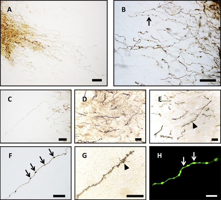
Figure 5.
Morphological characteristics of 11-month grafts suggest differentiation into multiple neuronal phenotypes. (A–H): Histological enhancement of green fluorescent protein-immunoreactive (GFP-ir) fibers revealed extensive fibritic growth reminiscent of mature monoamine neurons. Fine GFP-ir fibers adorned with varicosities (arrows) (B, F, H) projected within the substantia nigra (A) and were extremely dense at the ventro-caudal and dorsal-rostral region of the graft (B, C). Some GFP-ir processes were thicker and smooth (D) and may reflect nonterminal ends of axons, whereas other longer processes terminated in “spine-like” structures (arrowheads) (E, G). Scale bars = 250 μm (A), 50 μm (C, F, H), 25 μm (B, D, E), and 20 μm (G).

