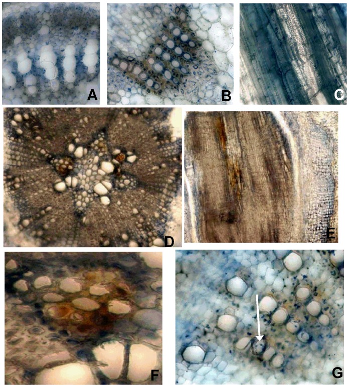
Figure 1. Latent infection in soybean stems infected with Phialophora gregata.
Latent infection as observed in stem cross sections from a BSR-susceptible soybean cultivar (Corsoy 79) infected with wild-type isolates of types A and B of Phialophora gregata. Images A–F were captured 2 weeks post inoculation. (A) No infection was observed in the cross sections of the apex (B) or the cross section and (C) longitudinal section of the middle of the non-inoculated control plant. (D) Necrosis in the vascular system of the root, (E) longitudinal section of a root, (F) a necrotic region from D that was cropped and magnified. Image G was captured 3 weeks post inoculation and hyphae) are beginning to colonize the xylem vessels (noted by arrow). Image A is 40x magnification, B, C, and E–G are 200x magnification, D is 100x magnification.

