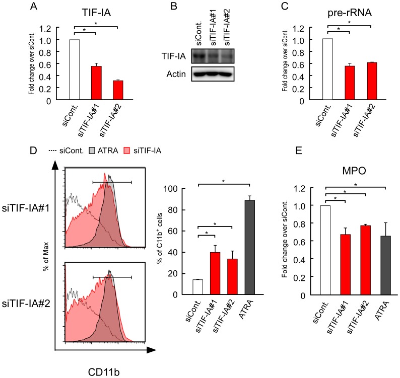
Figure 2. Suppression of rRNA transcription by TIF-IA KD induced the differentiation of HL-60.
(A, B) siRNA-TIF-IA reduced the mRNA and protein levels of TIF-IA. HL-60 cells were transfected with siRNAs for luciferase (siCont.) and TIF-IA (siTIF-IA#1 or siTIF-IA#2), and cultured for 3 days. (A) The mRNA levels of TIF-IA were determined by RT-qPCR. (B) The protein levels of TIF-IA were determined by immunoblotting. (C) The pre-rRNA levels were determined by RT-qPCR. (D, E) TIF-IA KD induced the differentiation of HL-60 cells. (D) CD11b expression was determined by flow cytometry. ATRA (1 µM) was used as the positive control. The corresponding mean percentages of CD11b-positive cells are shown in the left panels (right panels). We present the same histograms for siCont. and ATRA in the upper and lower panels because these experiments were performed at the same time. (E) The MPO levels were determined by RT-qPCR and normalized by the cyclophilin levels. Values are expressed as the mean ± S.D., n = 3 (A, B, D). Values are expressed as the mean ± S.D., n = 4 (C). *P<0.05.

