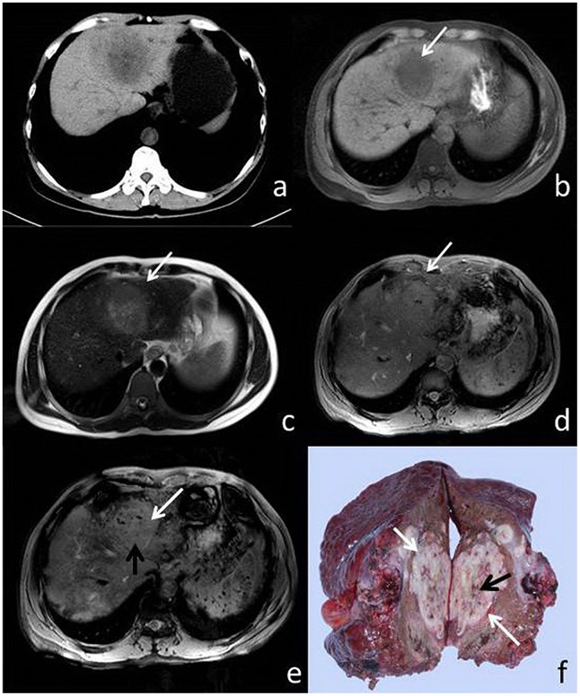
Figure 1. 51-year-old male with HCC.
(a) CT, (b) T1WI, (c) T2WI, (d) T2*, (e) SWI and (f) surgical specimen. The pseudocapsule is not perceptible on CT, T1W1, or T2WI (a–c), but is visible (white arrow) on both T2*WI and SWI (d, e). Both pseudocapsule and microhemorrhage (black arrow) are visible in the resected specimen (f) and SWI (e), and to a lesser extent, T2*WI (d).

