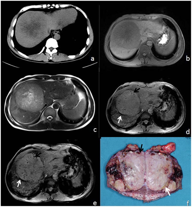
Figure 2. 65-year-old male with HCC.
(a) CT, (b) T1WI, (c) T2WI,(d) T2*, (e) SWI, and (f) surgical specimen. Pseudocapsule and foci of microhemorrhage are not visible on CT, T1WI, or T2WI (a–c), but the pseudocapsule (white arrow) and microbleeds (black arrow) are visible on both T2*WI (d) and SWI (e) as well as the surgical specimen (f). Here, SWI demonstrates the mosaic pattern of tumor heterogeneity (e).

