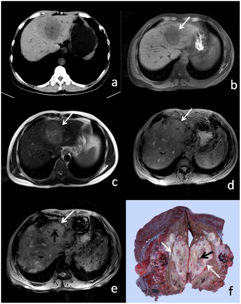
Figure 3. 46-year-old male patient with HCC.

(a) CT, (b) T1WI, (c) T2WI, and (d) T2* show no appreciable mosaic pattern or hemorrhagic foci. (e) SWI shows hypointense linear intratumoral structures (white arrow) and hemorrhagic foci (black arrow). (e) Resected surgical specimen shows internal microhemorrhage (white arrow) and linear structure (black arrow), which correlate with imaging findings.
