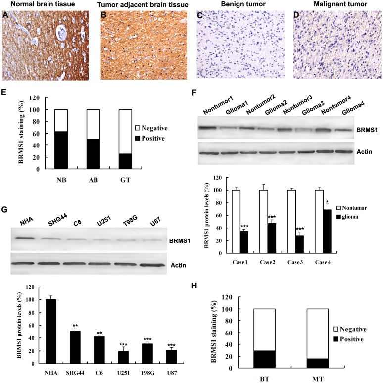
Figure 1. Representative images show BRMS1 immunohistochemical staining.
A Positive BRMS1 staining in normal brain tissue (NB); B Positive BRMS1 staining in adjacent normal brain tissue (AB); C Negative BRMS1 staining in benign tumor (BT); D Negative BRMS1 staining in malignant tumor (MT); E A significant difference in BRMS1 staining was observed between normal brain tissue and glioma tissue (GT) (P<0.01, χ2 test) and between tumor adjacent normal brain tissue and glioma tumor (P<0.01, χ2 test). F Whole-cell protein extracts were further prepared from four paired tumor adjacent normal glioma tissues and glioma tissues. The BRMS1 protein level was determined by Western blot analysis. G Western blot analysis of BRMS1 expression in normal human astrocytes NHA and glioma cell lines, including SHG44, C6, U251, T98G, U87. H BRMS1 staining was dramatically decreased in malignant tumor compared with benign tumor (P<0.05). All experiments were carried out in triplicate. Data are shown as mean ± SD. *P<0.05, **P<0.01, ***P<0.001. Original magnification (A–D) ×40.

