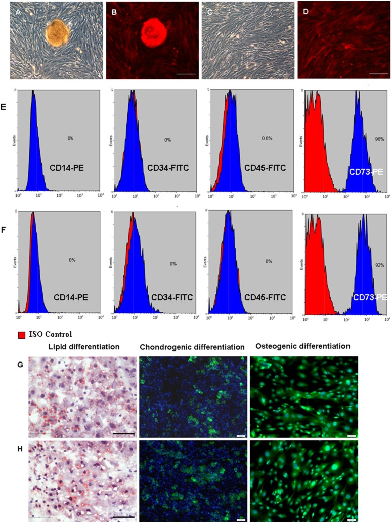
Figure 1. The characterization and differentiation of mouse SMSCs.
Morphology of A–B) F-SMSCs (a cell cluster in the middle of the dish) and C–D) M-SMSCs from RFP transgenic mice (scale bar, 200 µm). E) Phenotype of F-SMSCs by flow cytometry. Of the F-SMSCs, 96% expressed CD73 but not CD45, CD34, or CD14. F) M-SMSCs detected by flow cytometry. Of the M-SMSCs, 92% expressed CD73 but not CD45, CD34, or CD14. G) F-SMSCs and H) M-SMSCs were able to differentiate into adipocytes (oil red O staining), chondroblasts, and osteoblasts under standard in vitro differentiation conditions. The nuclei were counterstained with DAPI (blue) (scale bar, 50 µm).

