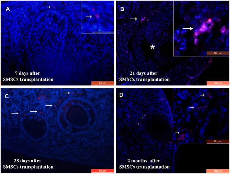
Figure 5. Transplantation of a line of RFP-transgenic SMSCs into chemotherapy-sterilized recipient mice.
A) Immunofluorescence of ovarian sections from sterilized mice 7 days after transplantation with RFP-transgenic SMSCs. Arrows indicate RFP staining in the ovarian stroma. B) RFP staining was observed around the antral follicle 21 days after transplantation. Asterisks indicate the antral follicle. C) RFP-positive cells were observed around granulosa cells in recipient ovaries 28 days after SMSC transplantation. D) RFP staining was observed in antral follicles of recipient ovaries 2 months after SMSC transplantation. Scale bars: A, C) 50 µm; B, D) 100 µm; A, insets) 20 µm; and B, D insets) 10 µm.

