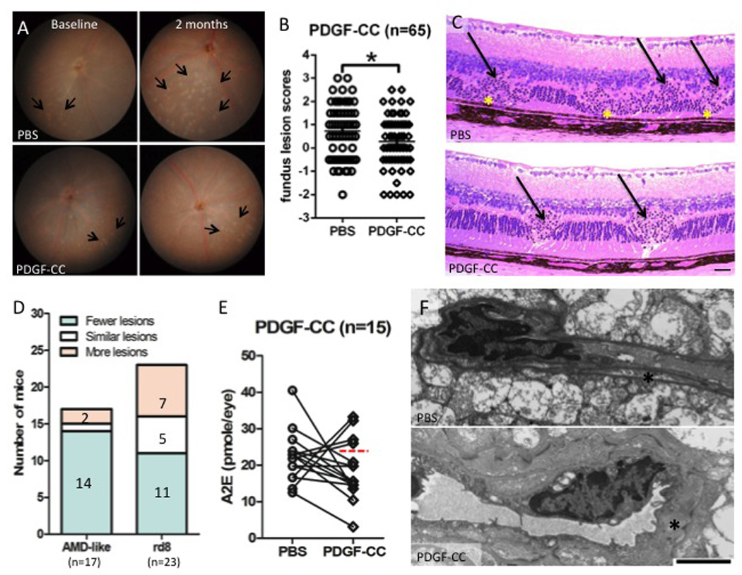
Figure 2. Retina-protective effect of PDGF-C in DKO rd8 mouse retina.

A) The fundus of PBS-injected and PDGF-CC-treated DKO rd8 mouse eyes was taken by a Nikon D90 digital camera illuminated with a Karl Storz veterinary otoendoscope. Representative fundus findings showed changes in yellowish deep retinal lesions (arrows) in both eyes of the same mouse before and 2 months after PDGF-CC treatment. B) Fundus scores of 65 pairs of eyes based on the fundus photographs showing altered progression scores between the two eyes at the end of 2 months after PDGF-CC treatment. C) DKO rd8 mouse eyes were enucleated and embedded in methacrylate. Representative histopathological findings showed retinal lesions of both DKO (asterisks) and rd8 (arrows) associated lesions. Scale bar = 50 µm. D) Histological comparison of AMD-like lesions and rd8-associated lesions between each mouse. The lesions in the PDGF-CC-treated eye are compared with that in PBS-injected eye of the same mouse. E) PBS-injected and PDGF-CC-treated DKO rd8 mouse eyes were enucleated and homogenized for A2E extraction with chloroform/methanol (n=15). The 11 eyes below the dashed line showed decreased A2E. F) DKO rd8 mouse eyes were enucleated and embedded with Spurr's epoxy resin. PDGF-C maintained pericytes (asterisks, scale bar = 2 µm) in retinal tissues when compared with PBS-injected eyes. *P<0.05.
