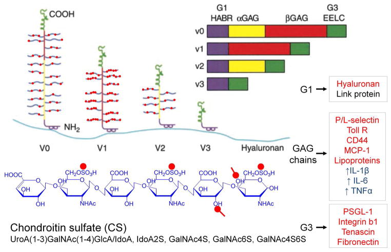
Figure 1.
A model of the different isoforms generated by alternative splicing of the mRNA transcript for versican. All isoforms interact with hyaluronan and thus are capable of forming different sized versican-HA aggregates. Different colors denote specific domains in the gene and in the protein product. Purple = hyaluronan binding region (HABR); yellow = α GAG exon and protein product; red = β GAG exon and protein product; green = two epidermal growth factor repeats (EE), a lectin binding domain (L) and a complement regulatory region. Inflammatory molecules that bind to different domains of versican are shown in the boxes to the right, marked in red. Those marked in blue indicate macrophage responses to treatment with versican. Structure of the CS GAG is shown at the bottom in blue with red dots denoting negatively charged residues. Reprinted (with modifications) from Current Opin Cell Biol, 14(5), Thomas N. Wight, Versican: a versatile extracellular matrix proteoglycan in cell biology, pp. 617–23, Copyright (2002), with permission from Elsevier.

