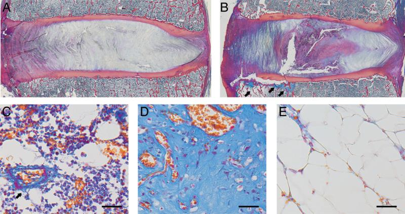
Figure 5.
(A) Mid-sagittal histology section from a spinal level with an intact cartilage endplate. (B) Endplates with damage were typified by cartilage avulsions and fissures at the interface between the inner annulus and nucleus pulposus. Note the fibrovascular and fatty marrow reactions adjacent to the location of the endplate damage (arrows). (C) Normal hematopoietic endplate marrow with blood vessel (arrow) and vascular sinusoids. (D) Richly vascularized endplate marrow with dense fibrous tissue corresponding to the arrows in panel B. (E) Fatty marrow with low cellularity corresponding to the leftmost arrow in panel B. In all panels, left side is anterior. In panels C–E, scale bars are 50 μm. Heidenhain connective tissue stain.

