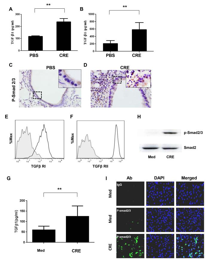
Figure 2.
Increased activation of TGFβ1signaling in lung tissue of allergic asthma and CRE-treated SCs. (A-B) Active TGFβ1 levels in BAL (A) and peripheral blood (B) of mice treated with saline and CRE were analyzed by ELISA. (C-D) Histological sections (p-Smad2/3 staining) of mouse airway treated with saline (C) and CRE (D). Data are representative of 3 independent experiments (A-D: n=4-6 mice/group). (E-F) Flow cytometry detected TβRI (E) and TβRII (F) expression in MSCs. (G) Active TGFβ1 levels in supernatants of CRE-treated (50μg/ml) and untreated MSCs for 72 hours. (H) Expression of p-Smad2/3+ in MSCs after exposed to CRE for 24 hours were analyzed by western blotting (H) and immunofluorescence analysis (I). Bars represent mean ± SEM of 3 independent experiments. **P<0.01.

