
Figure 5.
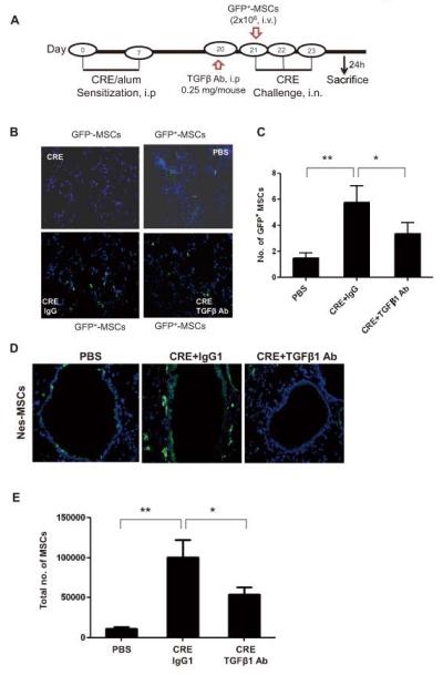
MSCs mobilize to the lungs from peripheral blood through TGFβ1. (A) Schematic of experimental protocol for mouse models of asthma. (B) Immunofluorescence analysis of injected GFP+MSCs in the airways of CRE-challenged or saline-treated mice with or without TGFβ1 Ab. (C) Number of injected GFP+MSCs were counted per field of view (FV, x20 magnification) and analyzed. Bars represent mean ± SEM for 4-6 mice/group. (D) GFP+ cells in the airways of CRE-challenged or saline-treated Nes-GFP mice. (E) Total numbers of MSCs in BAL detected by flow cytometry (anti-GFP). Bars represent mean ± SEM of 3 independent experiments. *P<0.05 **P<0.01.