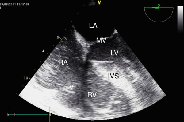
Figure 1.

Transoesophageal echocardiogram image demonstrating severe pulmonary hypertension and right ventricular overload with a dilated right ventricle and bulging of the interventricular septum to the left. Also visible is a dilated right atrium with spontaneous echo contrast. LA, left atrium; MV, mitral valve; LV, left ventricle; RA, right atrium; TV, tricuspid valve; RV, right ventricle; IVS, interventricular septum.
