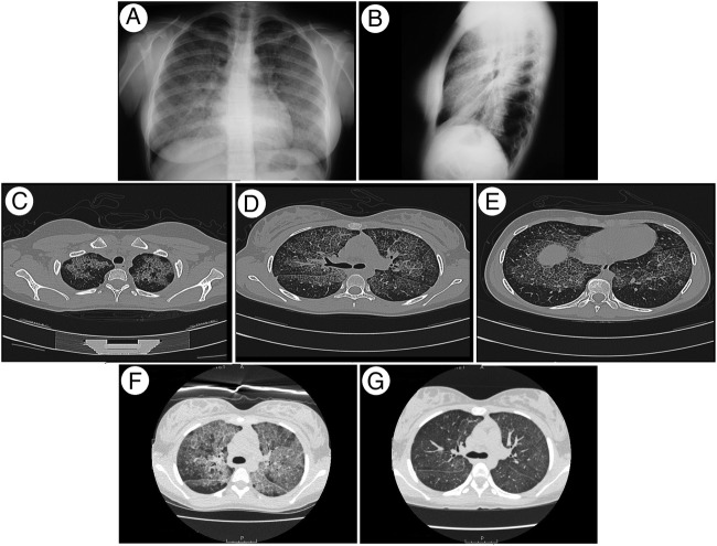
Figure 1.
Imaging studies. (A) Posteroanterior and (B) lateral chest radiographs showing widespread interstitial abnormalities, with a mid-zone and lower zone predominance and relative sparing of the apices and costophrenic angles. (C–E) High-resolution CT (HRCT) showing patchy geographic ground-glass opacities superimposed on interlobular septal thickening in multiple lobes (crazy-paving appearance). (F) Pre-WLL and (G) post-WLL HRCT showing a decrease in the density of the alveolar filling.

