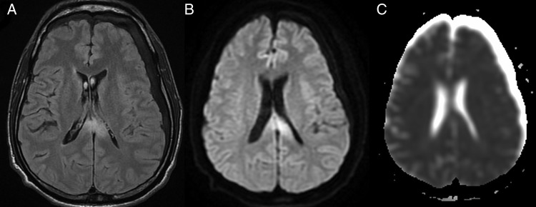
Figure 2.

(A, B) Follow-up MRI fluid attenuation inversion recovery (FLAIR) and diffusion images revealed persistent hyperintensity in the splenium. (C) Apparent diffusion coefficient images do not reveal corresponding hypointensity suggestive of T2 shine through. As compared with the prior MRI, there is a significant decrease in the extent of involvement.
